Please wait


Figures 1 Α-D. Α) Marking of slightly elevated early gastric cancer, type ‘0-IIa’ before endoscopic removal with ESD technique. Β) ESD ulcer bodem during ESD C) ESD gastric ulcer after ESD D) ESD specimen for histological diagnosis.

Figure 2. At the follow-up endoscopy one year after ESD removal normal scar was shown.

Figures 1 and 2 reffered to a 77-years-old male patient with early gastric cancer successfully diagnosed and treated by endoscopic curative rejection using ESD technique. ESD procedure in this case, was performed by Dr. Nikos Eleftheriadis, at the endoscopy Department of private clinic, under conscious sedation. Although the patient had a history of serious heart disease, ESD was successfully completed; patient mobilized the same day and discharged the next day after ESD, with gradual increase in diet from liquids to soft diet during a week after ESD. One year later patient was asymptomatic, while control endoscopy showed normal scar.

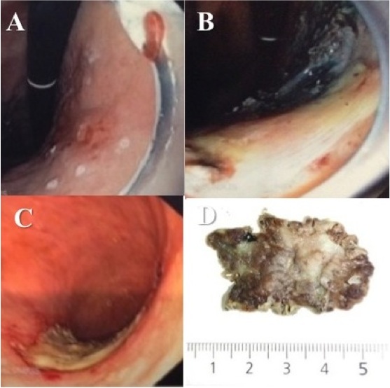
Figure 3 Α-D. Α) Sessile rectal adenoma (LST ‘0-IIa+ΙΙc’), 1cm from dentate line. Β) Submucosal space during rectal ESD C) ESD ulcer D) Rectal ESD specimen.
Figure 3 referred to a 65-years-old male patient with lower GI bleeding, due to sessile rectal adenoma, more than >4cm in size, close to dentate line, near anus. Patient underwent successful, curative endoscopic resection using ESD technique, performed by Dr. Νikos Eleftheriadis, at the endoscopy Department of private clinic, under conscious sedation. Rectal adenoma was completely removed in one specimen with normal margins (up to 5cm in diameter). Patient mobilized the same day and discharged next day. One year later he was totally normal, while control colonoscopy showed normal scar, avoiding permanent colostomy, which was the next alternative to ESD.


Figure 4 Α-D. Α) Submucosal neoplasm, ‘0-I’ on the ground of atrophic body gastritis. Β, C) ESD ulcer D) ESD specimen (3.2X2X0.3εκ)

Figure 5 Α-D. Α) ESD ulcer soon after procedure. Β)Haemostasis with coagulation forceps. C, D) ESD ulcer at the completion of ESD C) and next day D)Figures 4 and 5 referred to a 62-years-old male patient, with submucosal neoplasm, of the gastric body, up to 2 cm in size. Patient underwent successful curative endoscopic rejection, using ESD technique, performed by Dr. Nikos Eleftheriadis at the endoscopy Department of private clinic. The submucosal neoplasm was completely removed within normal margins in one piece, while histological examination showed carcinoid tumor (neuroendocrine tumor grade 1 (ΝΕΤ G1). No other further treatment was necessary.
Although the patient had a history of severe heart disease, and ESD was performed under anticoagulants subcutaneously, patient mobilized the same day, discharged next day, with gradual increase in diet from liquids to soft diet, during a week after ESD. During follow up one year later, patient is totally normal, control endoscopy showed normal scar, avoiding open surgery, which was the next alternative to ESD.

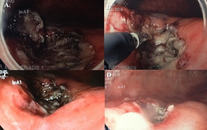
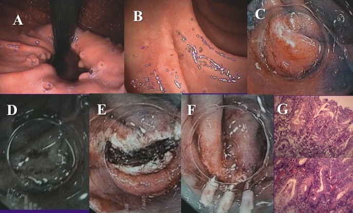
Figure 6 (A-G). A 30-years old male with FAP came for diagnostic gastroscopy. A) Multiple fundic gland gastric polyps were found. B) A small (0.5cm) depressed type 0-IIc nonampullary duodenal lesion was identified. C) Indigo carmine chromoendoscopy visualized better the lesion. D) NBI without magnification did not add more to WLE or indigo carmine chromoendoscopy. E-F) En bloc R0 resection followed by EMR-C and three clips (fig. F) were placed to close the EMR ulcer (fig. E). G) H&E stain of mucosectomy specimen shows depressed type duodenal adenoma (type III Vienna classification), with complete pathological rejection.


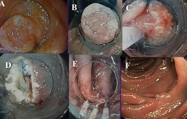
Figure 7 (A-F). A 60-years old male came for diagnostic gastroscopy. A) A small (0.5cm) depressed type 0-IIc nonampullary duodenal lesion was identified. B) Indigo carmine chromoendoscopy visualized better the lesion. C-E) En bloc R0 resection followed by EMR-C. Three clips (fig. E) were placed to close the EMR ulcer (fig. D). F) Histology showed duodenal adenoma (type III Vienna classification) with complete pathological resection. One year later a normal scar was identified and no residual tumor.

I am available to you all times to help and give solution to: ‘Achalasia’ with endoscopic myotomy - POEM technique, to reflux disease (GERD) and Barrett esophagus with antireflux mucosectomy (ARMS), to early gastrointestinal (GI) cancer (early cancer of the stomach and large bowel) with ESD technique, to Zenker diverticulum with myotomy (diverticulotomy) and in biliary diseases with ERCP.

Social Media

© 2018 Achalasia. All rights reserved- Created and hosted by Multihosting